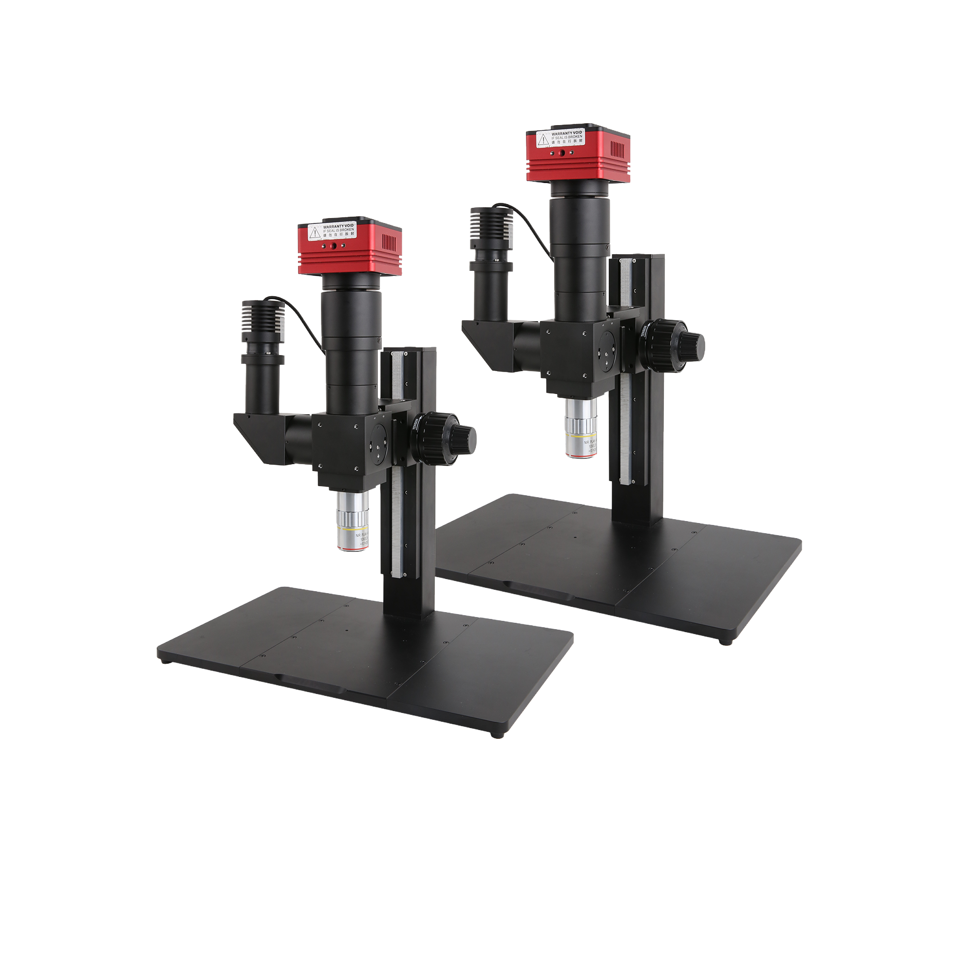
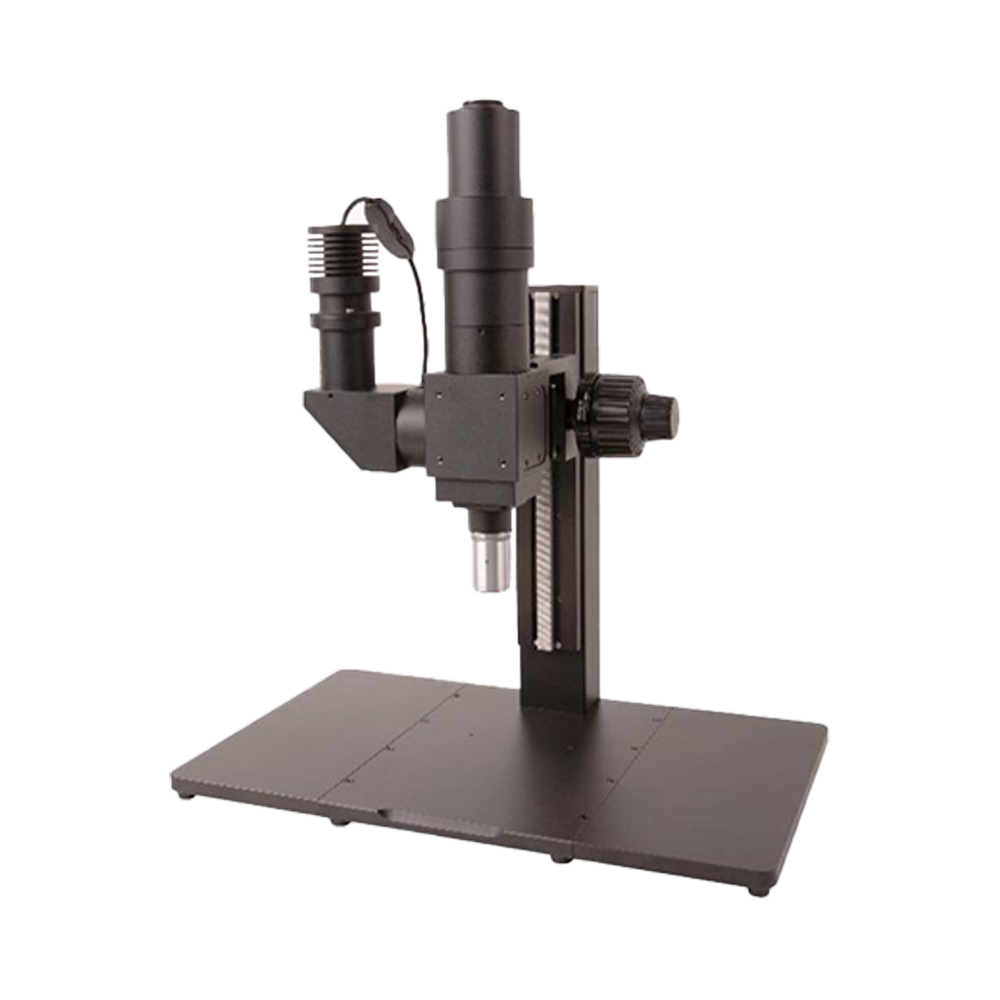
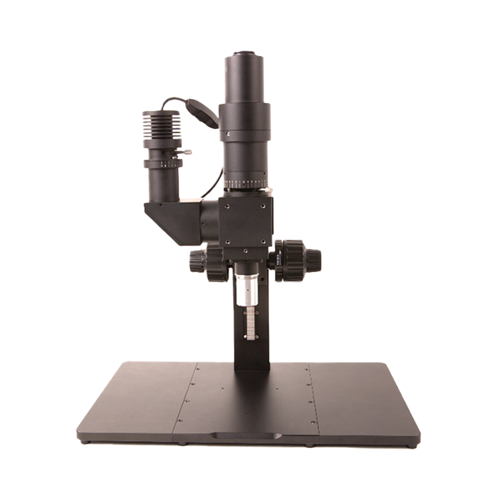
Specialized Microscopy Systems
Product Introduction
Professional microscopy systems designed for specialized application requirements, including short-wave infrared, differential interference contrast, brightfield metallographic, fluorescence, polarizing light, and other microscopy techniques.
Product Features
- Specialized design optimized for specific applications
- High-quality optical components
- Modular configuration for easy expansion
- Suitable for research and industrial inspection
- Complete system solutions
Find the right microscopy system for your application needs
Specialized Microscopy Systems
Professional microscopy systems designed for specialized application requirements, including short-wave infrared, differential interference contrast, brightfield metallographic, fluorescence, polarizing light, and other microscopy techniques.
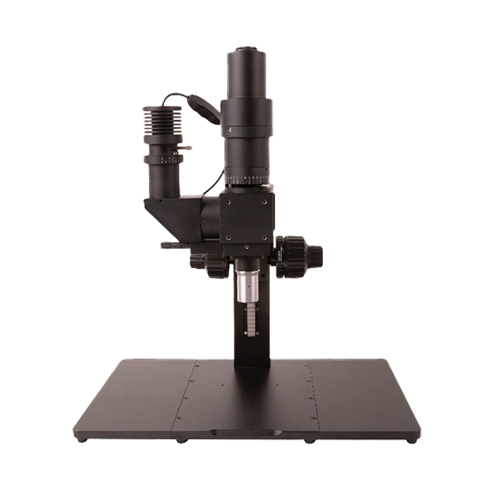
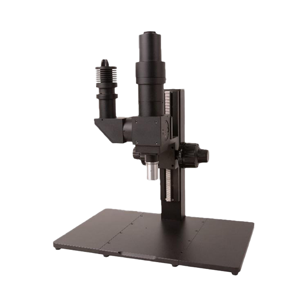
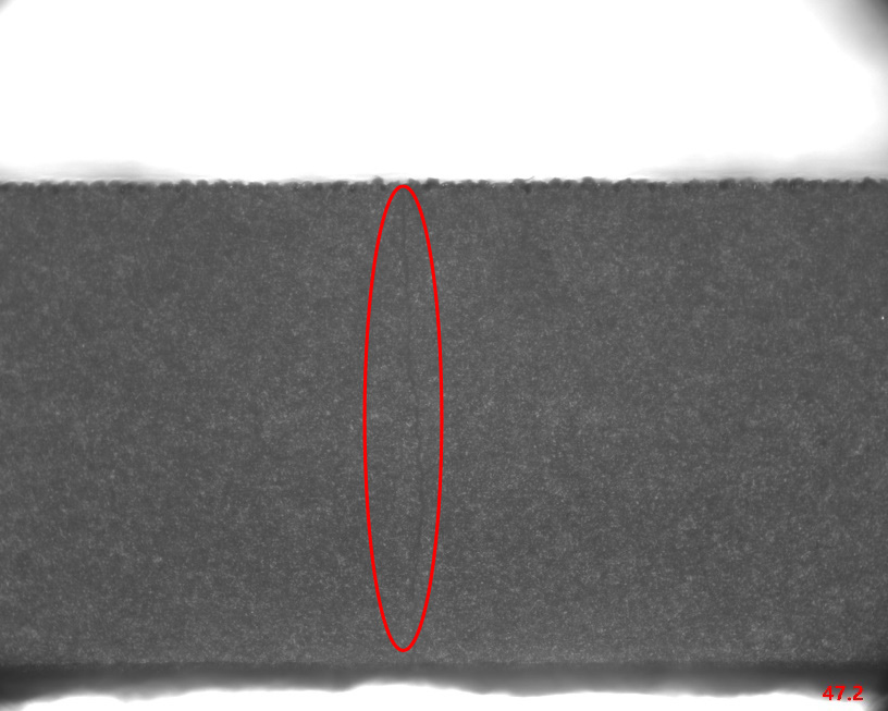
BSM - Short-Wave Infrared (SWIR) Microscopy System
The BSM Series modular SWIR microscopy system represents a new generation of Short-Wave Infrared imaging technology platform, extending the imaging ra...
Main Features
- Full coverage of 900-1700 nm SWIR imaging band
- Silicon material penetrative imaging technology for non-destructive internal inspection
- Three tube lens systems available (BSM-T180VA/T090VA/T100VA)
- Maximum 33 mm image field design, compatible with large-format sensors
- M Plan Apo NIR professional objective series (5X-50X HR)
- 0.4 µm ultra-high optical resolution (50X HR objective)
- Coaxial epi-illumination Köhler illumination system
- Integrated 1200/1300/1400/1550 nm multi-wavelength LED light sources
- Compatible with high-performance InGaAs sensor cameras (0.33M-5.0M)
- Real-time imaging capability with frame rates up to 400 fps@640×512
- TEC cooling technology ensuring low-noise, high SNR imaging
- Standard C-mount design compatible with various camera systems
- Modular architecture supporting flexible customization and upgrades
- Precision CNC machining with anti-vibration design
- Standard glass optical system for cost-effectiveness optimization
Application Fields
DIC100 - Differential Interference Contrast Microscopy System
DIC (Differential Interference Contrast) microscopy system utilizes the principle of dual-beam polarization interference, with the following process:
...
Standard Working Distance Objective Parameters (45 mm parfocal distance)
| Objective Name | Magnification | Numerical Aperture | Working Distance | Focal Length | Resolution | Object Field | Image Field | Thread Size |
|---|---|---|---|---|---|---|---|---|
| DIC2.5XA | 2.5X | 0.075 | 6.2 mm | 80 mm | 4.46 µm | 10 mm | 25 mm | M26×0.705 |
| DIC5XA | 5X | 0.15 | 23.5 mm | 39 mm | 2.2 µm | 5 mm | 25 mm | M26×0.705 |
| DIC10XA | 10X | 0.3 | 22.8 mm | 20 mm | 1.1 µm | 2.5 mm | 25 mm | M26×0.705 |
| DIC20XA | 20X | 0.4 | 19.2 mm | 10 mm | 0.8 µm | 1.1 mm | 25 mm | M26×0.705 |
| DIC50XA | 50X | 0.55 | 11 mm | 4 mm | 0.6 µm | 0.44 mm | 25 mm | M26×0.705 |
Long Working Distance Objective Parameters (60 mm parfocal distance)
| Objective Name | Magnification | Numerical Aperture | Working Distance | Focal Length | Resolution | Object Field | Image Field | Thread Size |
|---|---|---|---|---|---|---|---|---|
| DICL2XA | 2X | 0.055 | 33.7 mm | 100 mm | 6.1 µm | 12.5 mm | 25 mm | M26×0.705 |
| DICL5XA | 5X | 0.14 | 33.6 mm | 40 mm | 2.2 µm | 5 mm | 25 mm | M26×0.705 |
| DICL10XA | 10X | 0.28 | 33.4 mm | 20 mm | 1.2 µm | 2.5 mm | 25 mm | M26×0.705 |
| DICL20XA | 20X | 0.34 | 29.5 mm | 10 mm | 0.8 µm | 1.25 mm | 25 mm | M26×0.705 |
| DICL50XA | 50X | 0.5 | 18.9 mm | 4 mm | 0.7 µm | 0.5 mm | 25 mm | M26×0.705 |
Main Features
- Standard/Long working distance objective series (optional)
- Imaging optical path: 1X (tube lens focal length 180 mm), customizable with different magnification reducers
- Imaging path image field size: 25 mm
- Imaging path spectral range: Visible light
- Camera interface: C/M42/M52 optional
- Illumination method: Critical/Köhler illumination optional
- Illumination source: 10 W white/blue LED illumination optional
Application Fields
BMM100 - Brightfield Metallographic Microscopy System
The Brightfield Metallographic Microscope consists primarily of three major systems: illumination system, imaging system, and mechanical system. It is...
Standard Working Distance Objective Parameters (45 mm parfocal distance)
| Objective Name | Magnification | Numerical Aperture | Working Distance | Focal Length | Resolution | Object Field | Image Field | Thread Size |
|---|---|---|---|---|---|---|---|---|
| BF5XA | 5X | 0.15 | 22 mm | 36 mm | 2.23 µm | 5 mm | 25 mm | M20×0.705 |
| BF10XA | 10X | 0.3 | 15 mm | 18 mm | 1.1 µm | 2.5 mm | 25 mm | M20×0.705 |
| BF20XA | 20X | 0.4 | 10 mm | 9 mm | 0.75 µm | 1.25 mm | 25 mm | M20×0.705 |
| BF50XA | 50X | 0.8 | 2.5 mm | 3.6 mm | 0.41 µm | 0.5 mm | 25 mm | M20×0.705 |
Long Working Distance Objective Parameters (60 mm parfocal distance)
| Objective Name | Magnification | Numerical Aperture | Working Distance | Focal Length | Resolution | Object Field | Image Field | Thread Size |
|---|---|---|---|---|---|---|---|---|
| BFL2.5XA | 2.5X | 0.075 | 6.2 mm | 80 mm | 4.46 µm | 10 mm | 25 mm | M26×0.705 |
| BFL5XA | 5X | 0.15 | 23.5 mm | 40 mm | 2.2 µm | 5 mm | 25 mm | M26×0.705 |
| BFL10XA | 10X | 0.3 | 22.8 mm | 20 mm | 1.1 µm | 2.5 mm | 25 mm | M26×0.705 |
| BFL20XA | 20X | 0.4 | 19.2 mm | 10 mm | 0.8 µm | 1.1 mm | 25 mm | M26×0.705 |
| BFL50XA | 50X | 0.55 | 11 mm | 4 mm | 0.6 µm | 0.44 mm | 25 mm | M26×0.705 |
Main Features
- Standard/Long working distance objective series (optional)
- Imaging optical path: 1X (tube lens focal length 180 mm), customizable with different magnification reducers
- Imaging path image field size: 25 mm
- Imaging path spectral range: Visible light
- Camera interface: C/M42/M52 optional
- Illumination method: Critical/Köhler illumination optional
- Illumination source: 10 W white/blue LED illumination optional
Application Fields
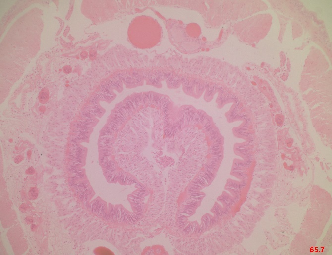
PLM100 - Professional Polarizing Light Microscopy Imaging Solution
The Polarizing Light Microscope is a microscope used for studying both transparent and opaque anisotropic materials. Any material with birefringence c...
Standard Working Distance Objective Parameters (45 mm parfocal distance)
| Objective Name | Magnification | Numerical Aperture | Working Distance | Focal Length | Resolution | Object Field | Image Field | Thread Size |
|---|---|---|---|---|---|---|---|---|
| POL2.5XA | 2.5X | 0.075 | 6.2 mm | 80 mm | 4.46 µm | 10 mm | 25 mm | M26×0.705 |
| POL5XA | 5X | 0.15 | 23.5 mm | 39 mm | 2.2 µm | 5 mm | 25 mm | M26×0.705 |
| POL10XA | 10X | 0.3 | 22.8 mm | 20 mm | 1.1 µm | 2.5 mm | 25 mm | M26×0.705 |
| POL20XA | 20X | 0.4 | 19.2 mm | 10 mm | 0.8 µm | 1.1 mm | 25 mm | M26×0.705 |
| POL50XA | 50X | 0.55 | 11 mm | 4 mm | 0.6 µm | 0.44 mm | 25 mm | M26×0.705 |
Long Working Distance Objective Parameters (60 mm parfocal distance)
| Objective Name | Magnification | Numerical Aperture | Working Distance | Focal Length | Resolution | Object Field | Image Field | Thread Size |
|---|---|---|---|---|---|---|---|---|
| POLL2XA | 2X | 0.055 | 33.7 mm | 100 mm | 6.1 µm | 12.5 mm | 25 mm | M26×0.705 |
| POLL5XA | 5X | 0.14 | 33.6 mm | 40 mm | 2.2 µm | 5 mm | 25 mm | M26×0.705 |
| POLL10XA | 10X | 0.28 | 33.4 mm | 20 mm | 1.2 µm | 2.5 mm | 25 mm | M26×0.705 |
| POLL20XA | 20X | 0.34 | 29.5 mm | 10 mm | 0.8 µm | 1.25 mm | 25 mm | M26×0.705 |
| POLL50XA | 50X | 0.5 | 18.9 mm | 4 mm | 0.7 µm | 0.5 mm | 25 mm | M26×0.705 |
Main Features
- Standard/Long working distance objective series (optional)
- Imaging optical path: 1X (tube lens focal length 180 mm), customizable with different magnification reducers
- Imaging path image field size: 25 mm
- Imaging path spectral range: Visible light
- Camera interface: C/M42/M52 optional
- Illumination method: Critical/Köhler illumination optional
- Illumination source: 10 W white/blue LED illumination optional
Application Fields
FM100 - Fluorescence Microscopy System
The Fluorescence Microscope is a microscope used for observing fluorescent or phosphorescent materials. The principle involves illuminating samples wi...
Standard Working Distance Objective Parameters (45 mm parfocal distance)
| Objective Name | Magnification | Numerical Aperture | Working Distance | Focal Length | Resolution | Object Field | Image Field | Thread Size |
|---|---|---|---|---|---|---|---|---|
| Flour2.5XA | 2.5X | 0.075 | 6.2 mm | 80 mm | 4.46 µm | 10 mm | 25 mm | M26×0.705 |
| Flour5XA | 5X | 0.15 | 23.5 mm | 39 mm | 2.2 µm | 5 mm | 25 mm | M26×0.705 |
| Flour10XA | 10X | 0.3 | 22.8 mm | 20 mm | 1.1 µm | 2.5 mm | 25 mm | M26×0.705 |
| Flour20XA | 20X | 0.4 | 19.2 mm | 10 mm | 0.8 µm | 1.1 mm | 25 mm | M26×0.705 |
| Flour50XA | 50X | 0.55 | 11 mm | 4 mm | 0.6 µm | 0.44 mm | 25 mm | M26×0.705 |
Long Working Distance Objective Parameters (60 mm parfocal distance)
| Objective Name | Magnification | Numerical Aperture | Working Distance | Focal Length | Resolution | Object Field | Image Field | Thread Size |
|---|---|---|---|---|---|---|---|---|
| FlourL2XA | 2X | 0.055 | 33.7 mm | 100 mm | 6.1 µm | 12.5 mm | 25 mm | M26×0.705 |
| FlourL5XA | 5X | 0.14 | 33.6 mm | 40 mm | 2.2 µm | 5 mm | 25 mm | M26×0.705 |
| FlourL10XA | 10X | 0.28 | 33.4 mm | 20 mm | 1.2 µm | 2.5 mm | 25 mm | M26×0.705 |
| FlourL20XA | 20X | 0.34 | 29.5 mm | 10 mm | 0.8 µm | 1.25 mm | 25 mm | M26×0.705 |
| FlourL50XA | 50X | 0.5 | 18.9 mm | 4 mm | 0.7 µm | 0.5 mm | 25 mm | M26×0.705 |
Main Features
- Standard/Long working distance objective series (optional)
- Imaging optical path: 1X (tube lens focal length 180 mm), customizable with different magnification reducers
- Imaging path image field size: 25 mm
- Imaging path spectral range: Visible light
- Camera interface: C/M42/M52 optional
- Illumination method: Critical/Köhler illumination optional
- Illumination source: 3 W power, 365 nm wavelength LED source
- Fluorescence module: DAPI single-band UV filter (excitation 365 nm, emission 445 nm, dichroic mirror 405 nm), customizable
Application Fields
Special Microscopy System Features
- Specialized design optimized for specific applications
- High-quality optical components
- Modular configuration for easy expansion
- Suitable for research and industrial inspection
- Complete system solutions
Selection Guide
Choose the appropriate microscopy method based on sample characteristics
Standard or long working distance objectives meet different needs
Köhler or critical illumination available
Supports C/M42/M52 and various interfaces
Topograph Photoelectric special microscopy systems adopt modular design, providing specialized solutions for different application scenarios to meet the high-end requirements of research and industrial inspection.
System Configuration Solutions
Flexibly combine hardware and software modules based on application requirements
| Dimension | Key Configuration | Technical Highlights | User Benefits |
|---|---|---|---|
| Imaging Hardware |
• ToupCam X Series: IMX415/IMX571 BSI CMOS, up to 45 MP, USB 3.0/HDMI 60 fps 4K • HCAM/PUM Portable Module: UVC plug-and-play, 8 LED ring light built-in |
• Low read noise & 66 dB+ dynamic range • Progressive scan + global shutter optional |
True color reproduction, high contrast; meets high-speed AOI, fluorescence weak signal and multi-scenario needs |
| Zoom Optics |
• MZO Series (0.25×–8×): 20× zoom ratio, NA 0.12, 174 mm long working distance • ZOPE All-in-one: Built-in 8 LED & USB camera, parfocal linear zoom |
Dual-end parallel optical path, diffraction-limited MTF, low distortion | Zoom without refocusing, view samples from millimeter to micrometer scale |
| Illumination System |
• TZM0756DRL 65/85 mm LED ring light: PWM continuously adjustable brightness • TZM0756CL coaxial light + point source • AALRL-200 large ring light: 300 mm uniform field |
Multi-channel/polarization/coaxial composite light; LED angle 30° adjustable | Solves PCB solder joint glare, wafer scratches, transparent film inspection challenges |
| Mechanical Platform |
• TPS-600 coarse/fine adjustment stand (5 kg load capacity) • TPS-300 precision fine adjustment 2 µm step • Motorized Z & XY platform (optional) |
Anodized Class II aerospace aluminum, ball screw | Long-term 24×7 stable positioning, supports autofocus and array scanning |
| Software & Algorithm |
• ToupView: Real-time measurement/annotation, deep focus synthesis, HDR, polarization demodulation • SDK/API: Windows/macOS/Linux/Android • AI Module: Defect classification, dimension tolerance judgment |
Secondary development + PLC/robot serial protocol | Quick integration into MES/SPC quality systems, supports edge computing and cloud synchronization |
System Advantages
Five core advantages building a professional microscopy imaging platform
Complete Ecosystem, Turnkey Delivery
All self-developed cameras, lenses, lighting, stands, and software — no multi-vendor procurement matching needed, plug-and-play saves 60% integration time.
High Resolution + Large Depth of Field
45 MP ultra-high definition CMOS + deep focus synthesis algorithm achieves micrometer-level depth clarity within a 30 mm field of view.
Multi-spectral & Low Light Imaging
Supports white light/near-infrared/polarization combination with coaxial + ring light synchronous exposure; reveals texture details even at 0.05 lux.
Flexible Expansion, Investment Protection
Standard C-Mount and GigE Vision/USB3 Vision protocols allow later upgrades to AI modules, motorized stages, and multi-camera synchronization without replacing the main unit.
Cross-Industry Implementation Cases
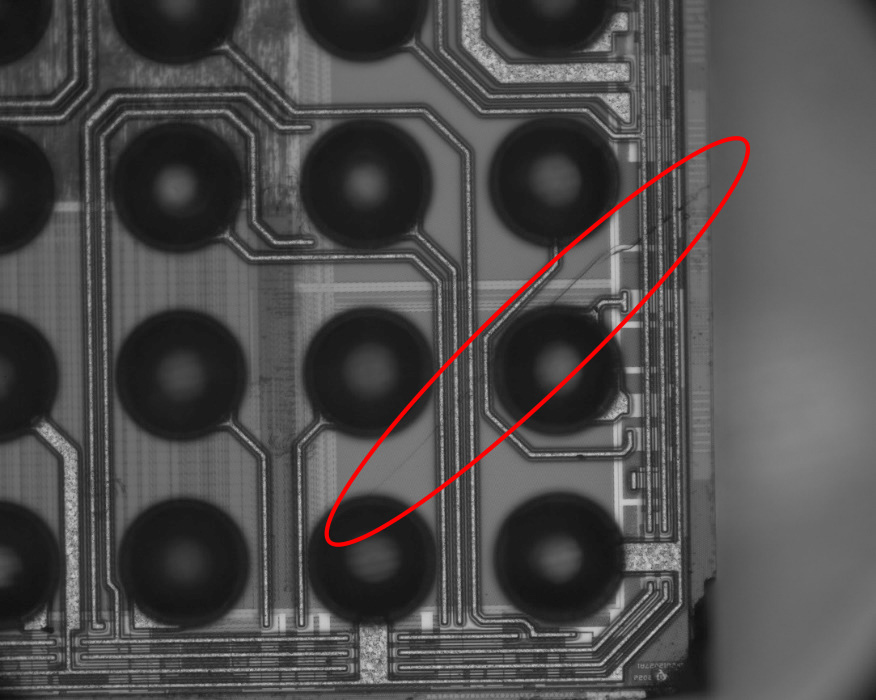
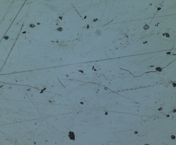
- Semiconductor: Bumping, scratch, wire bond defect AOI
- FPC/PCB: Solder paste height, pad residue detection
- New Energy: Lithium battery separator pore size, electrode coating consistency
- Life Sciences: Tissue sections, entomology, live plant observation
- Education & Training: University material science virtual experiments, STEAM maker courses
Application Cases
Successful implementation experience across multiple industries
Semiconductor Manufacturing
FPC/PCB Quality Control
New Energy Materials