Life Sciences
The application of the MTR3 series cameras in fluorescence microscopy
Areas of Application: Medical examinations, disease prevention, biological research,
educational and scientific research, etc.
-Routine fluorescence examinations in hospitals for fungi, gynecology, etc.
-Scientific research in universities and research institutes, alongside high-standard
diagnostics such as cancer confirmatory tests in hospitals.
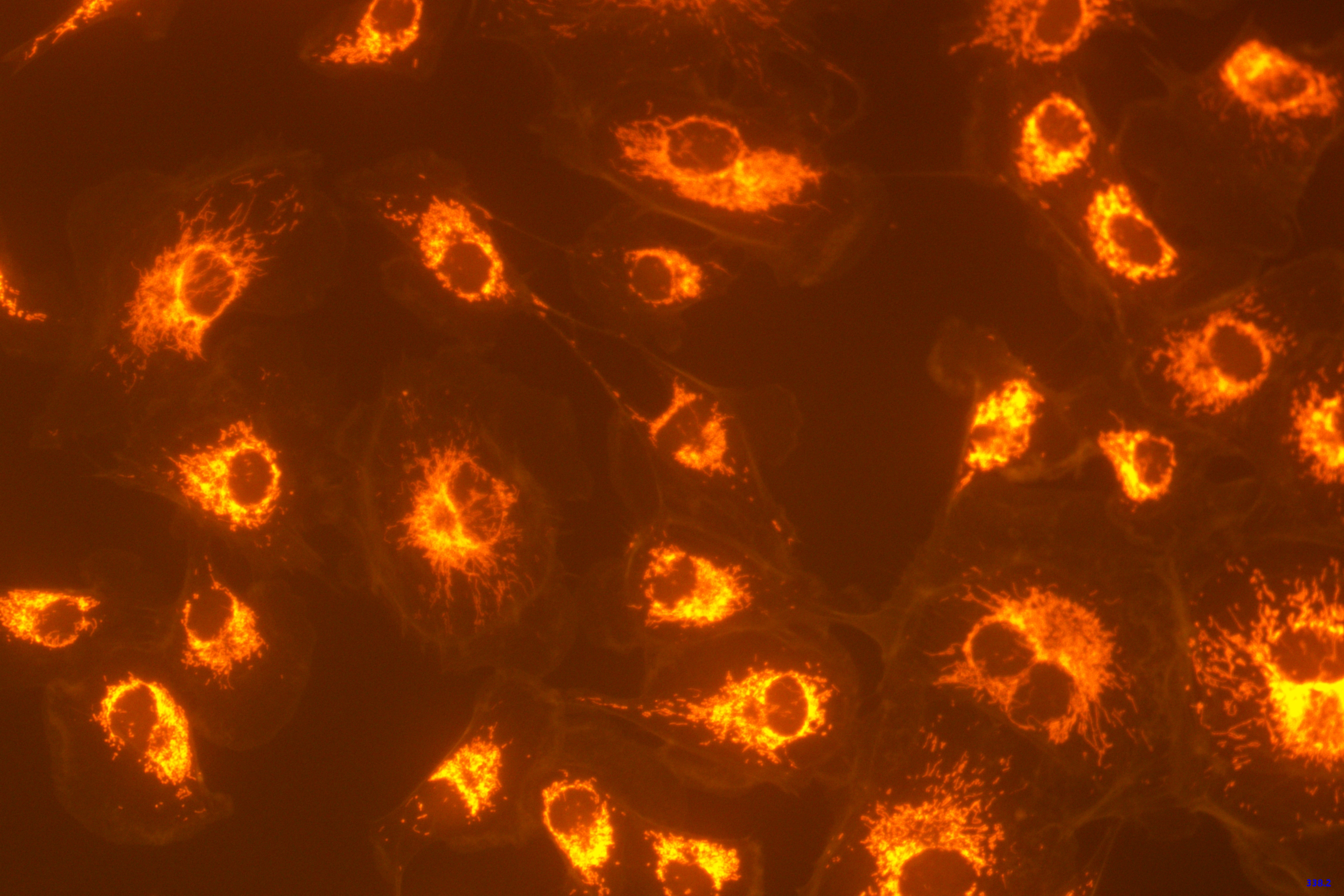
-Temporal observation of biological cells and other macroscopic fluorescence
observations.
Project Focus:
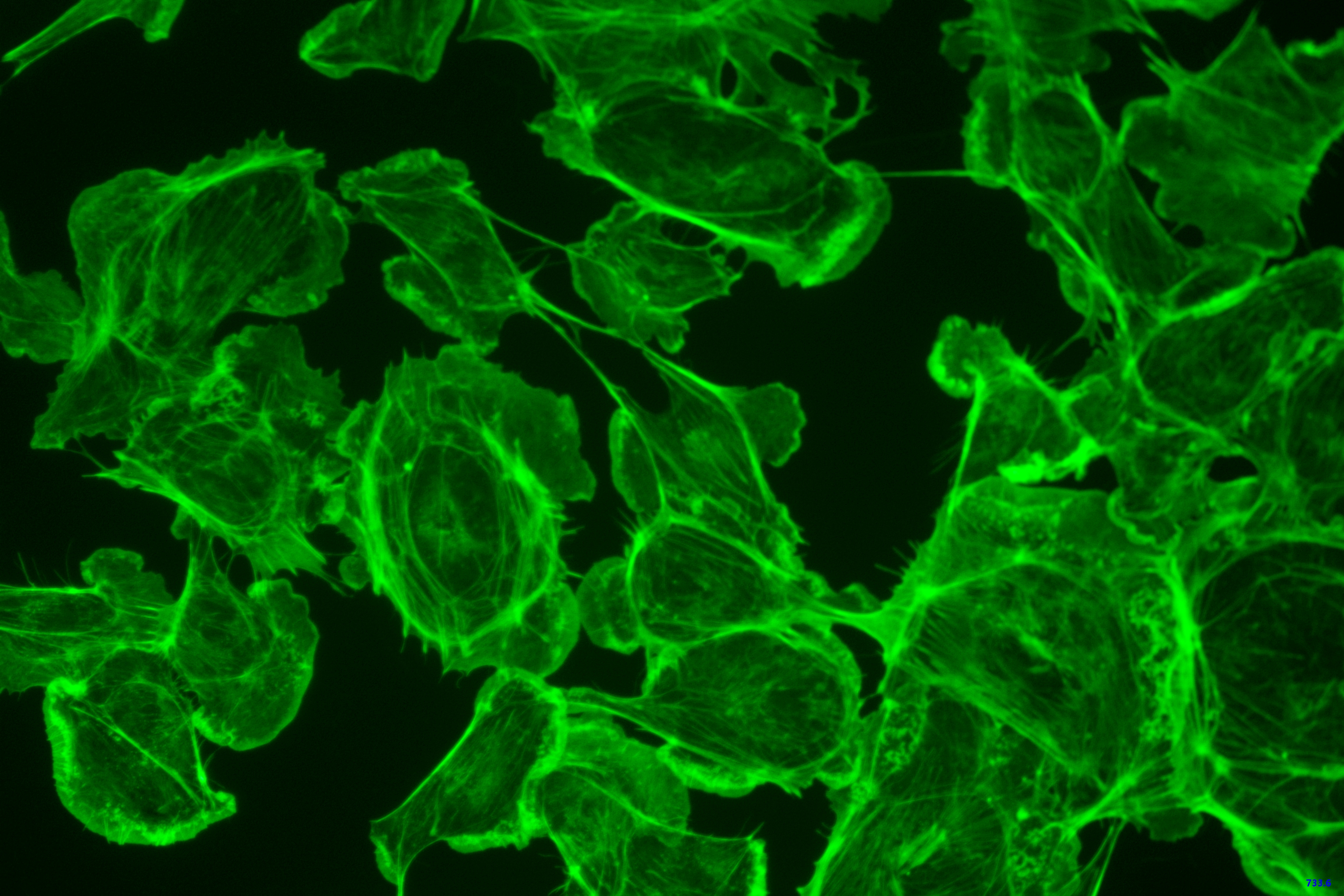
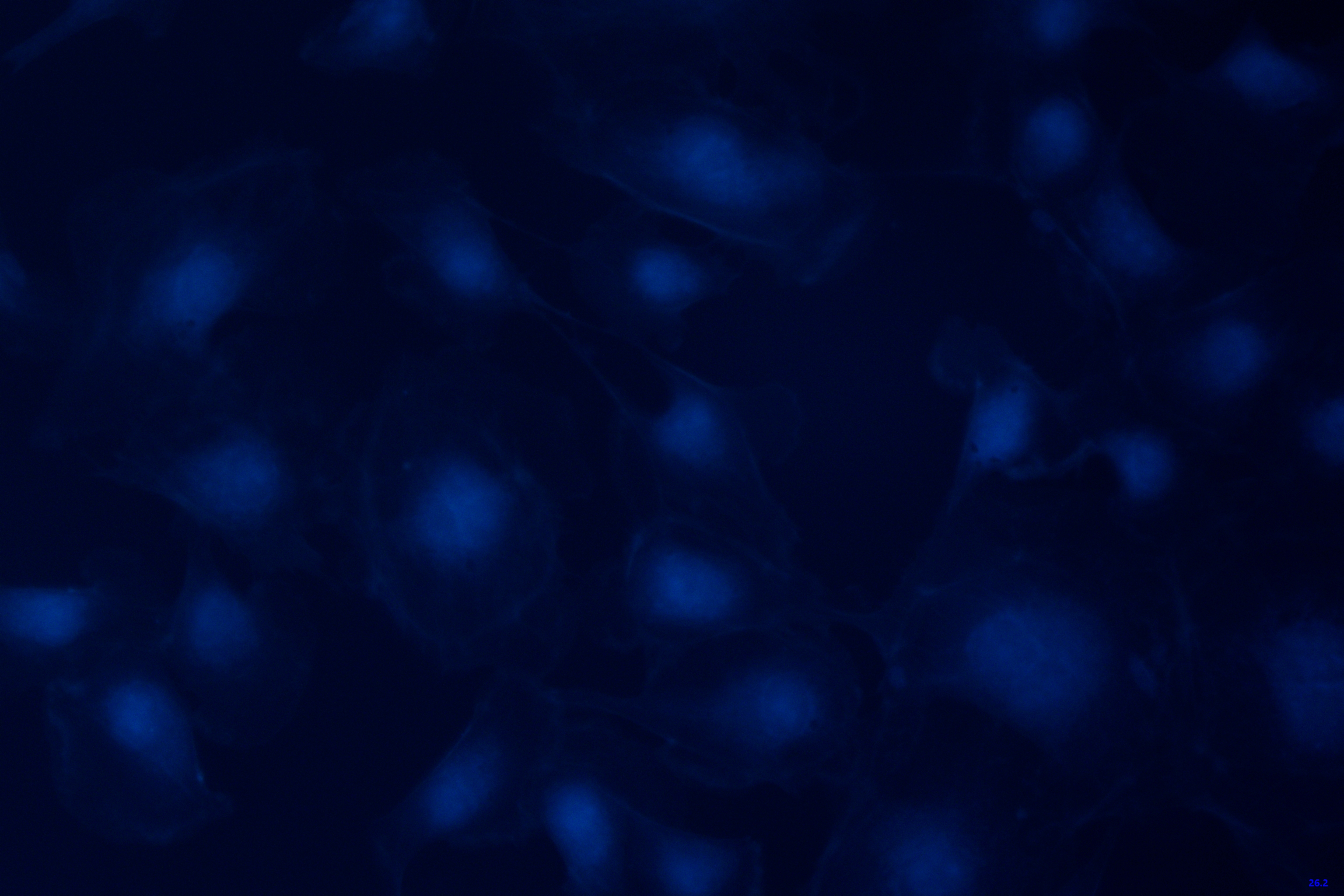
Fluorescence is a luminescence phenomenon generated by the transitions between internal
energy levels of molecules. When a substance, known as a fluorophore, absorbs light of a
specific wavelength (typically ultraviolet or blue light), it is excited to a higher
energy state. Upon returning to its ground state, the molecule emits light. The
wavelength of the emitted light is generally longer than that of the absorbed light,
implying that the energy of the emitted light is usually less than that of the absorbed
light. This occurs because a portion of the energy, when the molecule is excited to the
higher energy state, is lost in a non-radiative manner, resulting in the emitted light
having less energy than the absorbed light. This energy difference is also referred to
as the Stokes shift.
Fluorescence phenomena are categorized into two types: intrinsic fluorescence and
induced fluorescence.
Intrinsic Fluorescence: Certain substances naturally possess the property of generating
fluorescence. For example, certain organic molecules, minerals, or some biological
entities naturally absorb photons and release energy in the form of fluorescence.
However, in biological microscopy, intrinsic fluorescence often produces signals of
lower intensity and images with reduced contrast, which are generally undesired and may
hinder the observation of fluorescence-labeled samples due to the background of
intrinsic fluorescence.
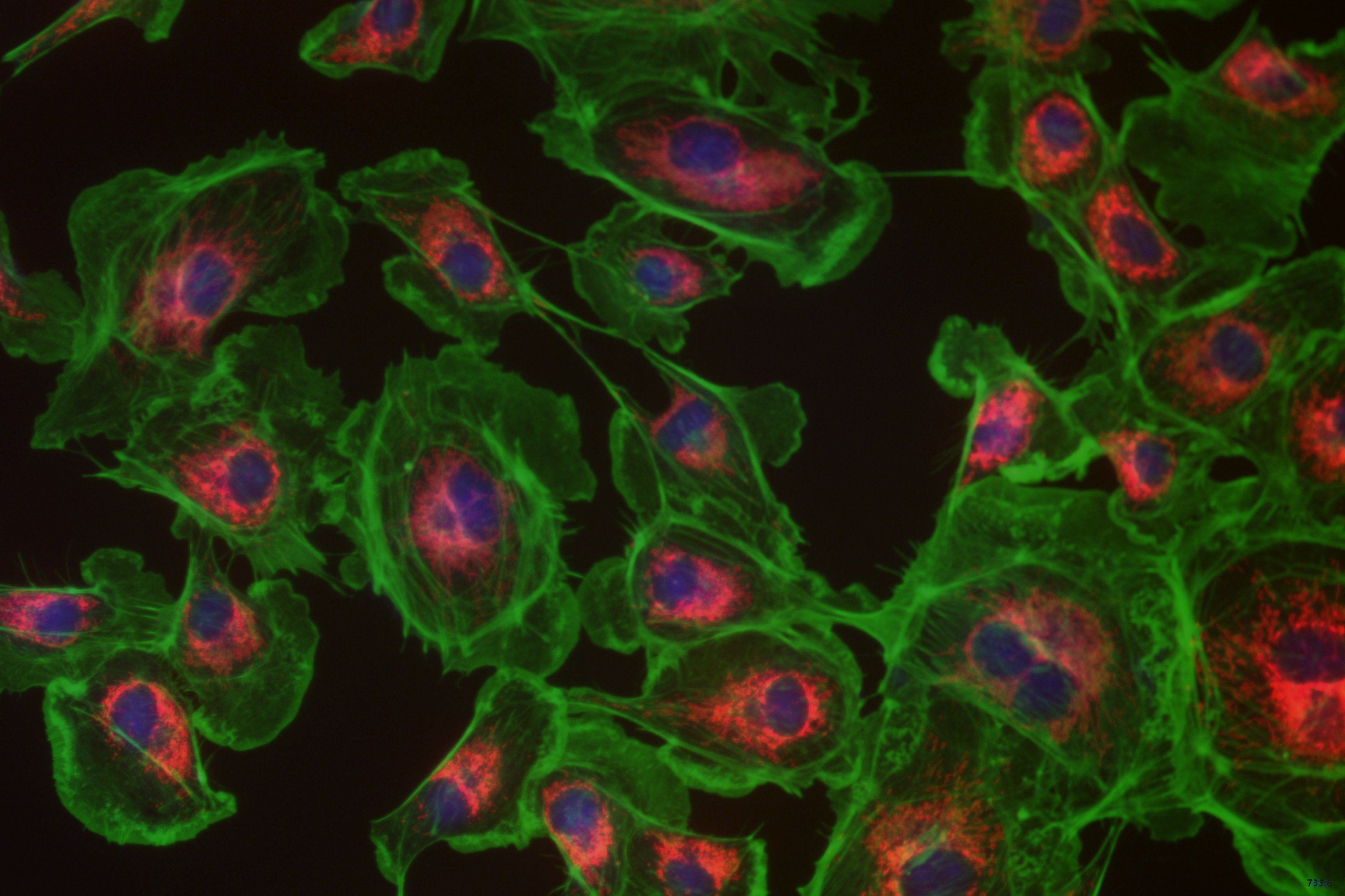
Induced Fluorescence: This involves the introduction of fluorescent dyes or labels into
a sample to enhance or induce fluorescence. Induced fluorescence, particularly through
secondary markers such as fluorescent proteins (like Green Fluorescent Protein, GFP) or
fluorescent probes (such as quantum dots or organic dyes), forms an essential component
of modern biological imaging and is widely utilized in biomedical research.
Case Synopsis:
In response to the burgeoning demands for precision and efficiency in cell culture
assays, the present case introduces a paradigm featuring high-definition spectral
imaging cameras, tailored image analysis algorithms, and an automated microscope
platform. This system employs a high-performance area array camera, boasting resolution
in the megapixel range, capable of capturing subtle variations in cellular structures
and is furnished with potent image processing software, facilitating real-time cellular
imaging and analysis. Through the intelligent visual guidance system, researchers are
empowered to automatically collect cellular images and apply advanced image processing
algorithms for cell counting, morphological analysis, and tracking the dynamic behaviors
of cells during the cultivation process. Moreover, the system is adept at identifying
abnormal cellular morphologies, serving as an early warning system for potential
cellular contamination or aberrant growth.
Advantages of the ToupTek MTR3CMOS Series:
- Offers a formidable resolution spectrum ranging from 500,000 to 45 million pixels;
- Features a dual-stage, professionally designed, highly efficient TE cooling
architecture that is ingeniously structured to facilitate rapid heat dissipation,
capable of precise temperature control with a maximum cooling reduction of 42 degrees
Celsius;
- Ingeniously designed anti-dew device ensures the sensor surface remains dew-free, even
in ultra-low temperatures;
- Offers R-CUT dual AR-coated protective glass as an optional accessory;
- Equipped with a high-speed USB 3.0 interface, achieving transfer speeds of up to
5Gbits/s;
- Equipped with a high-speed USB 3.0 interface, achieving transfer speeds of up to
5Gbits/s;
- Accommodates software or hardware trigger modes for capturing single or multiple
frames in videos;
- Utilizes an advanced Ultra-Fine color processing engine to ensure impeccable color
reproduction;
- Comes with advanced video and image processing application software;
- Provides a standard SDK compatible with multiple platforms including Windows, Linux,
macOS, and Android.
Introducing the advantages brought by the ToupTek MTR3CMOS series:
- Crisper and more detailed images
- Optimized light signal response allows for obtaining higher quality images even in dim
fluorescent conditions.
- Fast image capture for continuous recording of dynamic processes, reducing motion
blur.
-Enhanced dynamic range, crucial for analyzing differences in fluorescent signal
intensity.
-Reduced light exposure to samples, lowering the risk of photodamage to biological
specimens.
-Provides more accurate and consistent image capture, making data from repeated or
long-term experiments more reliable.
-High-quality images facilitate data sharing and collaboration among researchers,
improving research efficiency.
-ToupTek cameras offer advanced imaging functions at a more affordable price point,
greatly reducing costs.
ToupTek solemnly pledges to ensure the impeccable execution of each product and every
project. We provide our clients with the most rational product solutions tailored to their
needs.


