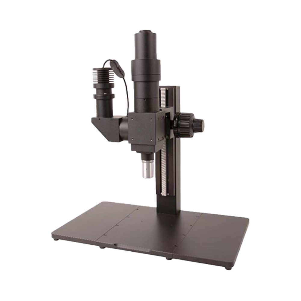
FM100 Series fluorescence microscope system
Product Introduction
A fluorescence microscope observes fluorescent or phosphorescent specimens. Excitation light of a specific wavelength (or band) illuminates the sample; fluorophores absorb the energy and emit longer-wavelength light. Emission filters separate the emitted light from excitation light so the detector records only fluorescence. In recent years, fluorescence microscopy has become indispensable for biological research, such as visualising fluorescently labeled biomolecules.
Product Features
- Standard and long working distance objectives available
- Imaging path: 1X (tube lens focal length 180 mm) with optional reducers
- Image circle: 25 mm
- Spectral range: visible light
- Camera interfaces: C / M42 / M52 (optional)
- Illumination: critical illumination or Köhler illumination (optional)
- Illumination source: 3 W, 365 nm LED
- Fluorescence module: DAPI single-band UV filter set (365 nm excitation, 445 nm emission, 405 nm dichroic mirror), customisable
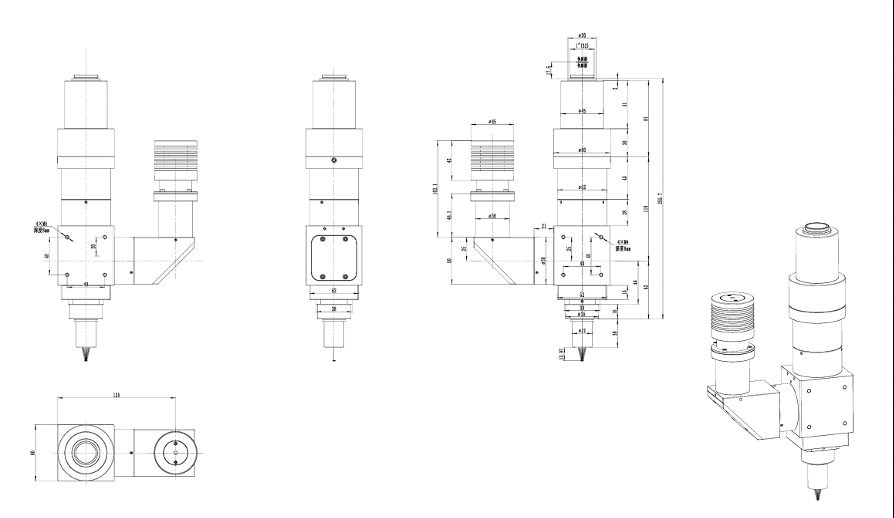
System Configuration and Parameters
Professional fluorescence microscopy system providing high-sensitivity imaging solutions for biomedical research and industrial inspection
System working principle
The FM100 fluorescence microscope system operates on fluorescence excitation and emission, using precision optical filters for selective signal detection.
Excitation illumination
Excitation light from the 365 nm UV LED is filtered to ensure only the target wavelength reaches the specimen.
Fluorophore excitation
Fluorophores in the sample absorb excitation energy, elevating electrons to the excited state.
Fluorescence emission
When excited electrons return to the ground state they emit longer-wavelength fluorescence (Stokes shift).
Signal separation
The dichroic mirror reflects excitation light and transmits emission light, while the emission filter further purifies the fluorescence signal.
Imaging inspection
Purified fluorescence signals are magnified by the objective and tube lens system before forming an image on the camera sensor.
Fluorescence System Configuration
Excitation Light Source
- Light Type
- UV-LED
- Power
- 3W
- Wavelength
- 365nm
- Lifetime
- >20000 hours
DAPI Filter Module
- Excitation
- 365 nm (bandwidth: 50 nm)
- Dichroic
- 405 nm
- Emission
- 445 nm (bandwidth: 50 nm)
- Suitable Dyes
- DAPI, Hoechst, etc.
Optical System
- Imaging Path
- 1X (180 mm tube lens)
- Image Size
- 25 mm
- Illumination
- Epi-fluorescence
- Uniformity
- >85%
Objective Series Parameters
Standard working-distance objective series (60 mm parfocal distance with a 200 mm tube lens )
| Model | Magnification | NA | Working Distance | Focal Length | Resolution | Object Field | Image Field |
|---|---|---|---|---|---|---|---|
| BF5XA | 5X | 0.15 | 23.5 mm | 39 mm | 2.2 µm | 5 mm | 25 mm |
| BF10XA | 10X | 0.3 | 22.8 mm | 20 mm | 1.1 µm | 2.5 mm | 25 mm |
| BF20XA | 20X | 0.4 | 19.2 mm | 10 mm | 0.8 µm | 1.1 mm | 25 mm |
| BF50XA | 50X | 0.55 | 11 mm | 4 mm | 0.6 µm | 0.44 mm | 25 mm |
Long working-distance objective series (95 mm parfocal distance with a 200 mm tube lens )
| Model | Magnification | NA | Working Distance | Focal Length | Resolution | Object Field | Image Field |
|---|---|---|---|---|---|---|---|
| BFL2XA | 2X | 0.055 | 33.7 mm | 100 mm | 6.1 µm | 12.5 mm | 25 mm |
| BFL5XA | 5X | 0.14 | 33.6 mm | 40 mm | 2.2 µm | 5 mm | 25 mm |
| BFL10XA | 10X | 0.28 | 33.4 mm | 20 mm | 1.2 µm | 2.5 mm | 25 mm |
| BFL20XA | 20X | 0.34 | 29.5 mm | 10 mm | 0.8 µm | 1.25 mm | 25 mm |
| BFL50XA | 50X | 0.5 | 18.9 mm | 4 mm | 0.7 µm | 0.5 mm | 25 mm |
Typical Application Cases
Professional applications of FM100 system in various fields

Biomedical Research
In biomedicine, the FM100 series plays a central role by using fluorescent labelling to precisely identify cellular and subcellular structures. It clearly visualises nuclei, cytoskeletons, and membranes, supporting cell biology research, pathology diagnostics, and drug discovery.
Case Parameters
- Highly specific labelling of target molecules
- Precise localisation of subcellular structures
- Simultaneous observation of multiple fluorescence channels
- Live-cell dynamic tracking
- Low-background, high-contrast imaging

Paper industry quality inspection
Within the paper industry, the FM100 series maps the distribution of fluorescent whitening agents. Under 365 nm UV excitation, the agents emit bright blue fluorescence, visualising otherwise invisible components and supporting quality control and formulation optimisation.
- Non-destructive inspection method
- Visualise bleaching agent distribution
- Quantitative analysis of fluorescent whitening agents
- Coating uniformity assessment
- Analysis of recycled paper composition
Other Application Fields
Materials science applications
In materials science, the system studies the fluorescence properties of polymers, composites, and nanomaterials, using fluorescent labelling to visualise internal structures, defect distributions, and stress states.
- Precise localisation of material defects
- Observation of polymer phase separation
- Nanoparticle dispersion evaluation
Food safety testing
The FM100 series supports rapid screening of natural fluorescing substances and additives in food—such as aflatoxins, vitamins, and preservatives—providing technical assurance for food safety supervision.
- Rapid toxin screening
- Additive composition analysis
- Microbial contamination detection
Forensic identification
In forensic science, the system examines body fluid traces, fibres, hair, and other evidence via fluorescence, using specific excitation wavelengths to enhance traces and support evidence collection.
- Enhanced visualisation of body fluid traces
- Fluorescent signature analysis of fibres
- Fingerprint fluorescence enhancement
Fluorescence imaging considerations
Avoid excessive fixation during sample preparation to prevent autofluorescence.
Select appropriate fluorophores to match excitation and emission wavelengths
Control excitation intensity to prevent photobleaching
Use anti-quenching reagents to extend fluorescence observation time
Operate in a darkroom to improve imaging contrast
Regularly calibrate filter combinations to maintain optimal performance.
Advantages compared with other microscopy techniques
| Comparison Technology | FM100 System Advantages |
|---|---|
| DIC microscopy | FM100 offers extremely high detection sensitivity and specificity, capable of capturing single-molecule fluorescence signals. |
| Phase-contrast microscope | FM100 supports multiplex labelling and molecular-specific identification rather than relying solely on refractive-index differences. |
| Darkfield microscope | FM100 delivers higher signal-to-noise ratio and quantitative analysis performance, enabling fluorescence intensity quantification. |
| Confocal microscope | FM100 features a simple, cost-effective architecture suited to routine fluorescence observation and wide-field imaging. |
System Configuration and Accessories
Standard Configuration
- FM100 main system
- 3 W 365 nm UV-LED light source
- DAPI fluorescence filter module
- Precision focusing stage
- UV-protective goggles
Optional Accessories
- Standard and long working distance objective sets
- Rotating specimen stage
- FITC fluorescence filter module
- TRITC fluorescence filter module
- Multi-position filter turret
- Automated stage control
- Image acquisition and analysis software
- Critical/Köhler illumination module
- Fluorescence reference standard slide
The FM100 Series supports customization of various fluorescence filter modules to meet observation requirements for different fluorescent dyes
FM100 System Advantages
Professional fluorescence microscopy technology achieving high-sensitivity specific imaging
Ultra-High Detection Sensitivity
Capable of detecting single-molecule level fluorescence signals, enabling visualization of extremely low concentration target substances.
Molecular-Specific Recognition
Achieves precise localization and quantitative analysis of specific molecules through fluorescent labeling technology.
Multiple Labeling Capability
Supports simultaneous observation of multiple fluorescent dyes, enabling co-localization analysis of multi-target molecules.
LED Cold Light Technology
3 W high-power UV-LED light source with low heat generation, service life exceeding 20,000 hours, and low operating costs.
Optimized Filter Design
High-quality DAPI filter module with high excitation efficiency, low background noise, providing excellent signal-to-noise ratio.
Wide-Field Imaging Advantage
Large field of view observation capability, suitable for overall imaging of large samples such as tissue sections and cell cultures.